Processing and Visualization of Biomedical Imagery
The remarkable achievements of life sciences over the last decade in monitoring, detection, and treatment of various diseases have lead to even higher expectations by the society in the future. Although new sensing modalities are continuously being developed, followed by an ever increasing stream of biomedical data, the analysis capabilities by humans have not changed. Effective tools for analyzing, representing and visualizing the captured data are of essence for the future of patient diagnosis and treatment.
Efforts of the VIP lab in this area have concentrated on both model-based and machine-learning techniques for the quantitative analysis and classification of biomedical imagery based on metallomic, photometric and other image attributes, and also on enhanced visualization using 3-D stereoscopic and multiview displays.
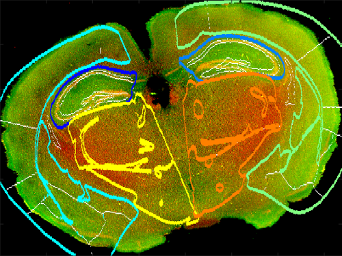
- Quantitative Analysis of Metallomic Images to Assess Traumatic Brain Injury: Novel image processing algorithms to quantitatively determine the link between TBI severity and CTE neuropathology
- Automatic Scoring of Bowel Preparation from Colonoscopic Imagery: Application of machine learning techniques to colonoscopy video classification.
- 3-D Automultiscopic Displays in the Treatment of Motor Functions and Balance: Development of 3-D displays for the treatment of bed-ridden patients and astronauts.
